Unquestionably! We should separate the data about the natural eye into various segments:
Definition:
The natural eye is an organ that identifies light and is the essential organ of vision in people. It works by capturing and processing visual information, allowing individuals to perceive their surroundings.
Anatomy:
The human eye has various anatomical components:
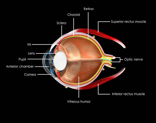
Cornea:
- The cornea is the straightforward forward portion of the eye that covers the iris, pupil, and front chamber.
- It plays a crucial role in focusing light into the eye and enabling clear vision.
- The cornea is composed of several layers, and its transparency is essential for maintaining the clarity of vision.
Sclera:
- The sclera is the intense, white, external layer of the eye that offers underlying help and security.
- It is commonly referred to as the “white of the eye.”
- The sclera, along with the cornea, forms the external protective coat of the eyeball.
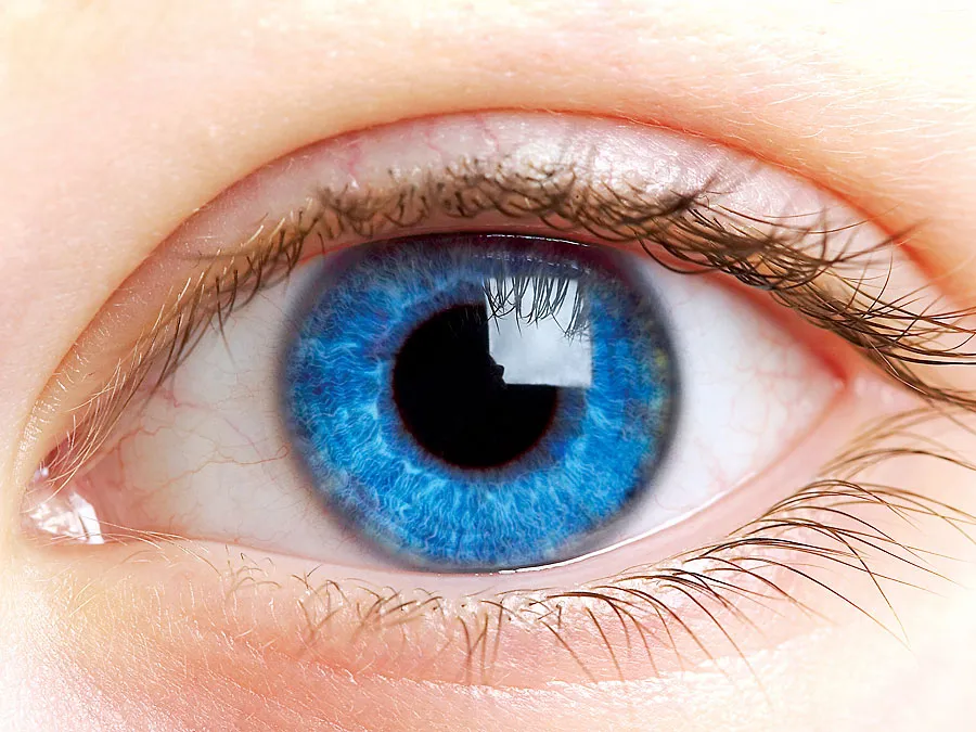
Iris:
- The iris is the shaded piece of the eye that encompasses the student, and it is situated between the cornea and the focal point.
- The iris plays a crucial role in regulating the amount of light that enters the eye, contributing to the eye’s overall visual function.
Pupil:
- A black circular opening in the center of the iris adjusts to control light entry.
Lens:
- The transparent, flexible structure behind the iris further focuses light.
The focal point is a straightforward, biconvex construction in the eye that assists with shining light onto the retina. It is situated behind the iris and changes shape to adjust the focal length, allowing the eye to focus on objects at varying distances.
Retina:
- The retina is a perplexing and sensitive layer of tissue situated at the rear of the eye.
- It is a crucial component of the visual system and plays a key role in the process of vision.
- The complex structure and functions of the retina make it a critical component in the visual process.
- Any abnormalities or damage to the retina can affect vision, emphasizing the importance of regular eye examinations for maintaining eye health.
Macula:
- The macula is a small, highly specialized area located in the central part of the retina at the back of the eye.
- It is crucial for central vision, which is essential for activities such as reading, driving, and recognizing faces.
- The macula contains a high centralization of cone cells, which are responsible for nitty-gritty vision and variety insight.
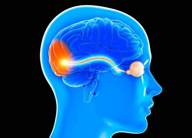
Optic Nerve:
- The optic nerve is a heap of nerve filaments that communicates visual data from the retina to the cerebrum.
- It plays a crucial role in the visual system by carrying electrical signals generated by the photoreceptor cells in the retina to the brain, where they are interpreted as visual images.
Vitreous Humor:
- The vitreous humor is a gel-like substance that fills the posterior part of the eye, occupying the space between the lens and the retina.
Aqueous Humor:
- The aqueous humor is a clear, watery fluid that fills the anterior (front) and posterior (back) chambers of the eye, between the lens and the cornea.
- It plays a crucial role in maintaining the eye’s shape, nourishing the cornea and lens, and helping with intraocular pressure.
Conjunctiva:
- The conjunctiva is a slender, straightforward film that covers the front surface of the eye (with the exception of the cornea) and lines the internal surface of the eyelids.
- It assumes a few significant parts in safeguarding and greasing up the eye.
Eyelids and Eyelashes:
- Eyelids protect the eyes; eyelashes prevent foreign objects.
Eyelid and eyelash problems can be caused by various factors, including infections, inflammatory conditions, allergic reactions, mechanical issues, etc.
Diagram:
A diagram can visually represent the structure of the human eye, showing the relationship between its different parts and their functions.
Function:
The primary function of the human eye is to capture and process visual information. Light enters through the cornea and lens, forming an inverted image on the retina. Photoreceptor cells in the retina convert this image into electrical signals, which are transmitted via the optic nerve to the brain. The brain then interprets these signals, allowing us to see and perceive the visual world.
Facts:
The human eye is capable of perceiving a vast range of colors.
Both eyes work together to provide depth perception.
Blinking helps spread tears across the surface of the eye, keeping it moist.
The eye is sensitive to light and adjusts through the dilation and constriction of the pupil.
The human eye is an intricate and sensitive organ, requiring care and protection.
Understanding the anatomy and function of the human eye is crucial for maintaining eye health and addressing vision-related issues. Regular eye exams are essential for the early detection and management of eye conditions.
Internal link: opticalsworld