


The eye is a complex organ responsible for vision, allowing us to perceive and interpret the surrounding world. Here are the main parts of the eye and an overview of how vision works:
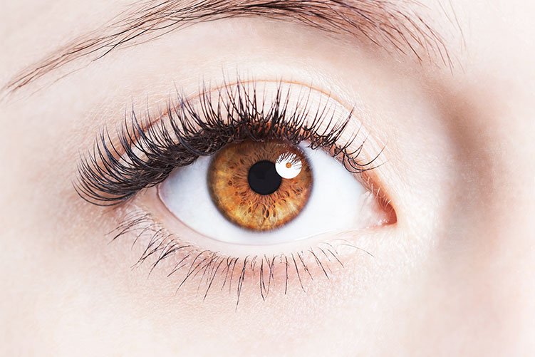
Cornea:

The cornea is the obvious the front a part of the attention that covers the iris, pupil, and anterior chamber.Along with the anterior chamber and lens, the cornea refracts light, accounting for about two-thirds of the eye’s overall optical power.In humans, the refractive energy of the cornea is about forty three dioptres.The cornea may be reshaped with the aid of using surgical approaches together with LASIK. While the cornea contributes maximum of the eye’s focusing power, its awareness is fixed. Accommodation (the refocusing of mild to higher view close to objects) is completed via way of means of converting the geometry of the lens.Medical phrases associated with the cornea frequently begin with the prefix “kerat-” from the Greek phrase κέρας, horn.
Iris:
The iris is the coloured a part of the attention surrounding the pupil.It controls the quantity of mild getting into the attention via way of means of adjusting the dimensions of the pupil.In maximum mammals and birds, the iris (pl.: irides or irises) is a thin, annular form withinside the eye, answerable for controlling theDiameter and length of the pupil, and consequently the quantity of mild achieving the retina. Eye colour is described through the iris.In optical terms, the student is the eye’s aperture, even as the iris is the diaphragm.
Pupil:
The scholar is a hollow placed withinside the middle of the iris of the attention. That lets in mild to strike the retina. It seems black due to the fact mild rays getting into the student. Are both absorbed with the aid of using the tissues internal theEye directly. Or absorbed after diffuse reflections inside the attention that in most cases leave out exiting the slim pupil.[citation needed] The length of the student is managed through the iris, and varies dependingOn many factors, the maximum widespread being the quantity of mild withinside the environment.The term “pupil” become coined with the aid of using Gerard of Cremona. In humans, the student is circular, however its form varies among species; a few cats, reptiles, and foxes Have vertical slit pupils, goats and sheep have horizontally orientated pupils, and a few catfish have annular types. In optical terms, the anatomical scholar is the eye’s aperture and the iris is the aperture stop.The photo of the scholar as visible from outdoor the attention is the doorway scholar, which does notprecisely correspond to the region and length of the bodily student due to the fact it’s miles magnified with the aid of using the cornea.On the internal aspect lies a distinguished structure, the collarette, marking the junction of the embryonic pupillary membrane masking the embryonic pupil.
Lens:
The lens is a transparent, bendy shape positioned in the back of the iris. It focuses light onto the retina by changing its shape through a process called accommodation.
Retina:

The retina is a light-sensitive layer at the back of the eye, similar to the film in a camera. It consists of cells referred to as photoreceptors—rods and cones—that convert mild into electric signals.
Rods and Cones:
Rods and cones are types of photoreceptor cells in the retina. Rods are responsible for vision in low light conditions. While cones are responsible for color vision and detailed vision in well-lit conditions.
Optic Nerve:
The optic nerve is a package deal of nerve fibers that consists of visible records. From the retina to the brain.
Vitreous Humor:
The vitreous humor is a clear, gel-like substance that fills the gap among the lens and the retina. Assisting preserve the form of the eye.
How We See:
Light Entry:
Light enters the eye through the cornea and passes through the pupil.
Lens Refraction:
The lens focuses the incoming light onto the retina by changing its shape through accommodation.
Image Formation:
The focused light forms an inverted image on the retina.
Photoreceptor Activation:
Photoreceptor cells (rods and cones) in the retina are activated by the light, converting it into electrical signals.
Signal Transmission:
The electrical signals are transmitted through the optic nerve to the brain.
Brain Interpretation:
The brain interprets the signals received from the optic nerve, forming the visual perception of the surrounding environment.
The process of vision involves the intricate coordination of these eye structures. And the brain, allowing us to perceive the world around us with depth, color, and detail.
Internal link : opticalsworld